
Oshika T, Inamura M, Inoue Y, et al 4 ABSTRACT SUMMARY Incidence and Outcomes of Repositioning Surgery to Correct Misalignment of Toric Intraocular Lenses
#ITRACE TORIC LENS ALIGNMENT SOFTWARE#
The AS-OCT software evaluated in this study holds promise for improving results with toric IOLs.


Equally important is the ability to troubleshoot when results are less than optimal.

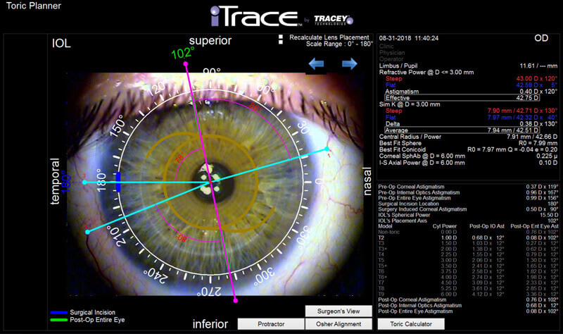
The emphasis (among both ophthalmologists and patients) on precision and accuracy in cataract and refractive surgery continues to grow. That said, the investigators noted that limits to the IOL software include a necessity for good visualization of IOL marks, which may not be possible in an eye with a small pupil, corneal opacities, or anterior capsular fibrosis. Additionally, images are acquired and assessed automatically, independent of the operator’s skill, and they have a resolution power of 1º, affording high interobserver repeatability. 1 Lucisano and colleagues used new toric IOL AS-OCT software that allows simultaneous analyses of corneal topography and the anterior segment in a single, rapid scan without the need to reposition the patient. 1-3 Drawbacks to these techniques are that they depend partially on subjective judgment, require a multistep analysis, and do not take into account the position of the patient’s head or cyclotorsion during fixation-all of which can lead to measurement variability. Surgeons have used several techniques to assess the postoperative alignment of a toric IOL (eg, slit-lamp examination, digital overlay, computer analysis of retroilluminated photographs). They recorded identical IOL rotation with the patient’s head tilted right and left, thus maintaining the values of misalignment found in primary position. The researchers found that, with the head in primary position, mean IOL misalignment from the intended topographic steep axis was 1.4º ☑.5º. Lucisano and colleagues evaluated postoperative IOL rotation in 15 eyes of nine patients. Rotation of the toric IOL from the intended position translated as the difference in degrees between the topographic axis and the value calculated from the linear marker. A screen layout created after the scan showed a topographic map on one side and an anterior segment image on the other together, they produced an image of the anterior segment with an overlapping green linear marker that could be rotated on a pivot centered over the corneal apex. Prior to the scan, the examiner tilted the patient’s head slightly to the right and left to determine the influence of head position on IOL position. The procedure involved a single, quick (< 0.3 seconds), noncontact, noninvasive, 3D scan while the patient was seated in primary position. The investigators dilated the pupil in order to visualize the marking dots in the periphery of the toric IOL. The new technique is easy to perform and less subjective than other methods. There are no standard algorithms for assessing IOL position postoperatively, and many methods are subjective and variable among examiners. Misalignment of the IOL may be the cause of residual refractive error after the implantation of a toric lens.


 0 kommentar(er)
